-
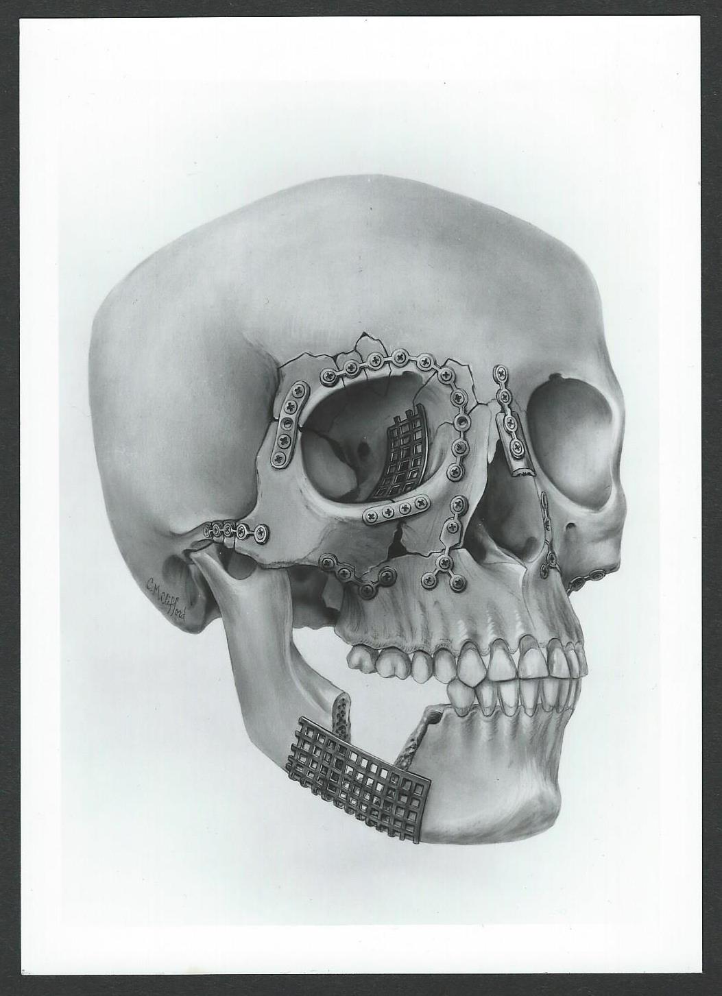
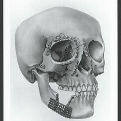
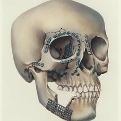
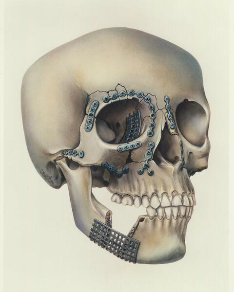
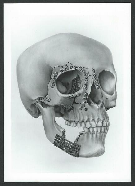 Plating of facial fracturesCarbon dust on calcium board. I drew this from a human skull that I had plated with glued-on photocopies of the plates. I penciled the fractures directly onto the skull, so that I could create various fracture and repair patterns by erasing and repositioning plates.
 -
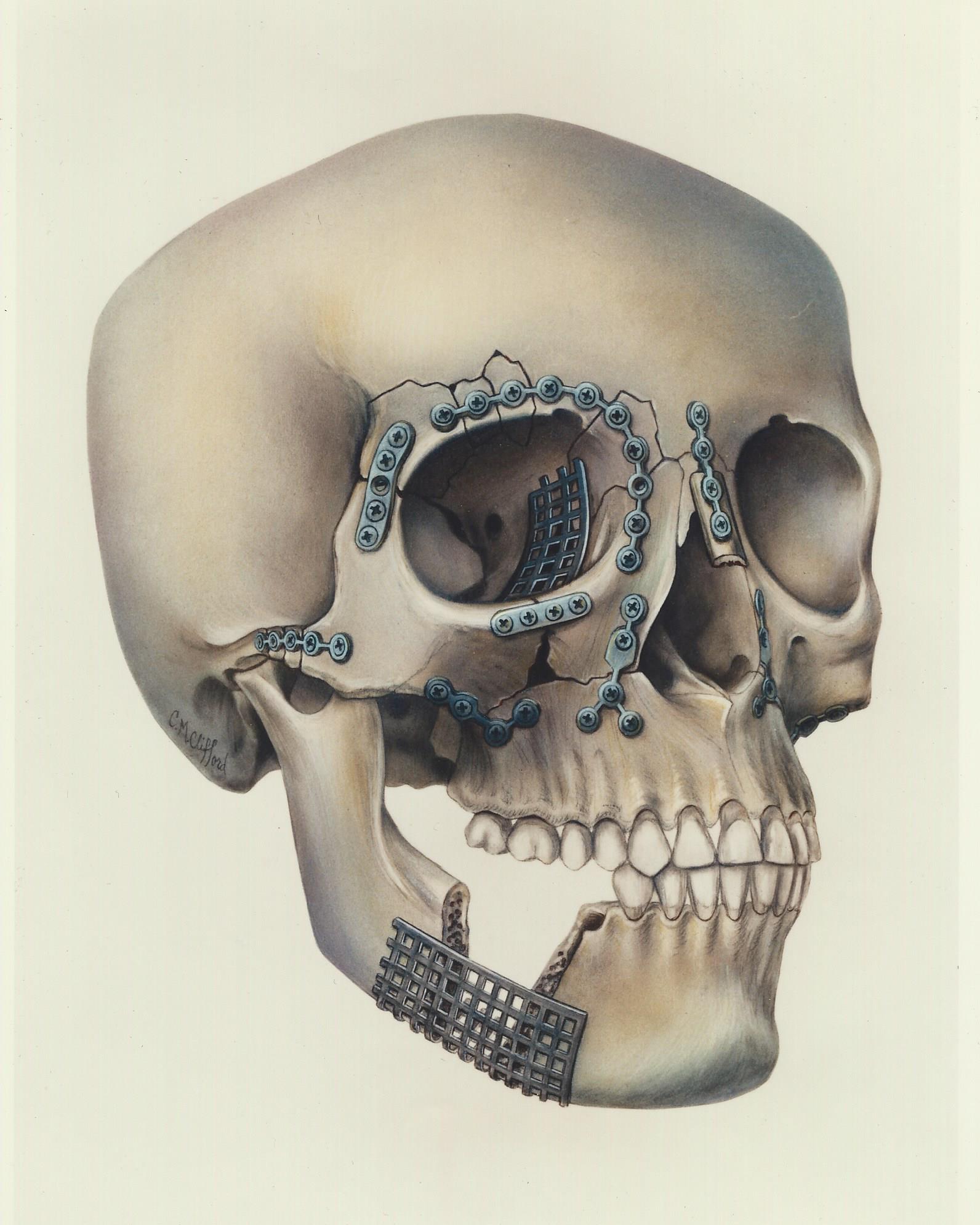
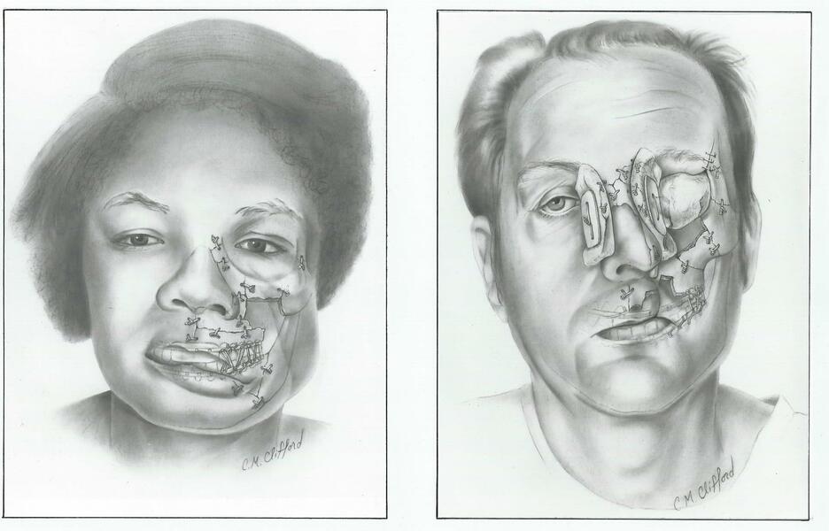
 Plating of facial fracturesAirbrush on photo paper. I made an extremely light print of the previous illustration and airbrushed it to make it full color.
 -
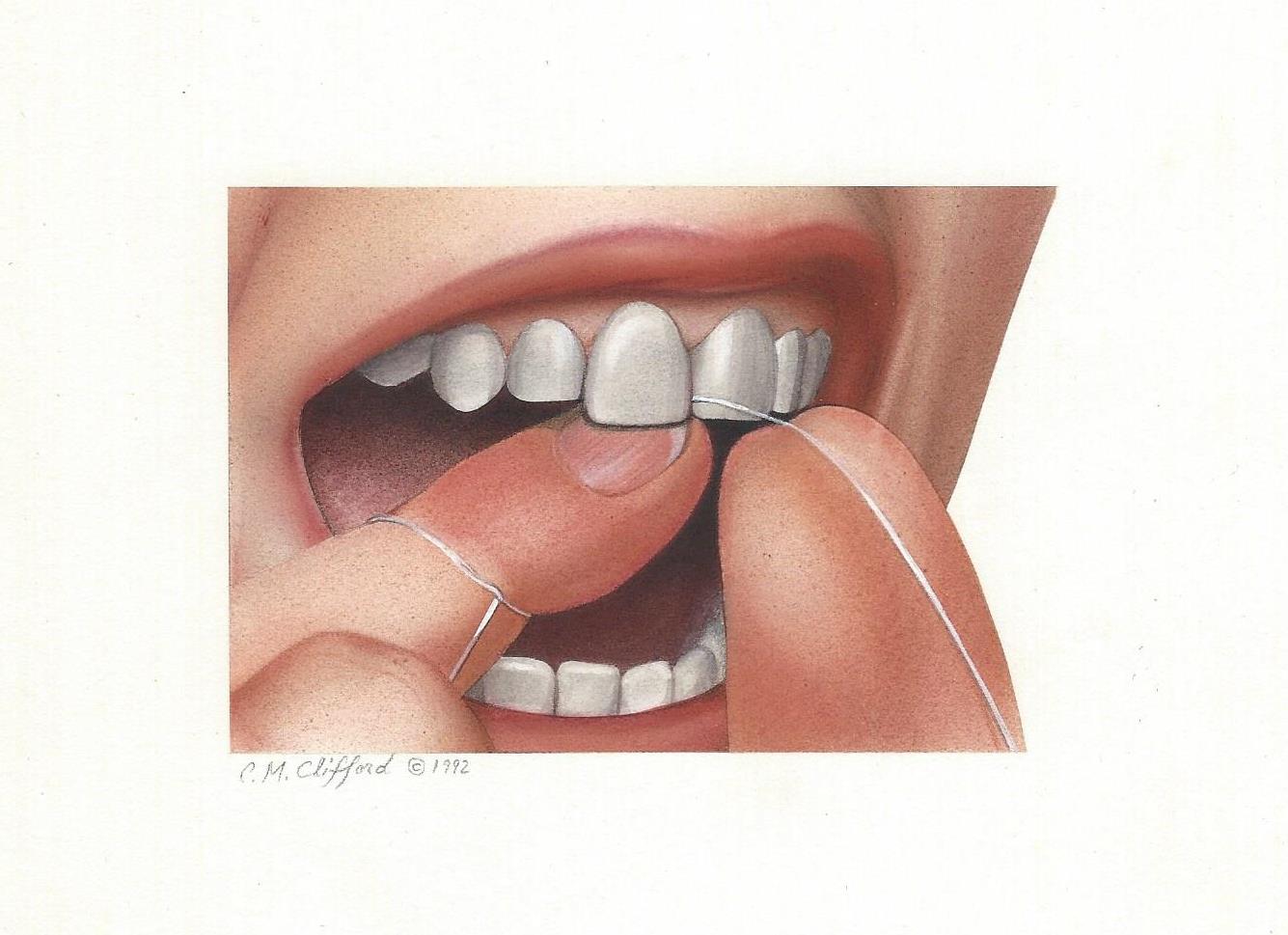
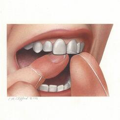
 How to FlossAirbrush on illustration board. 3 x 4 1/2 inches. Part of a series on how to floss.
 -
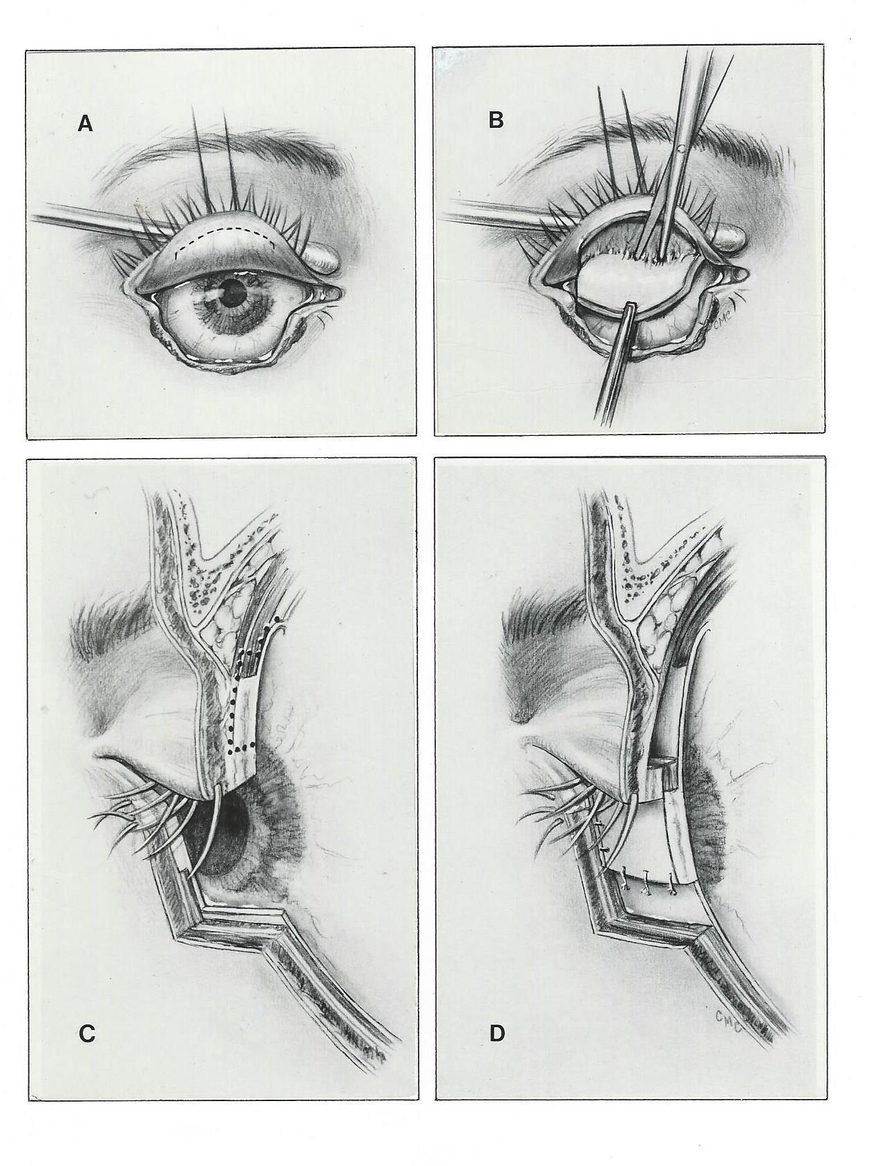
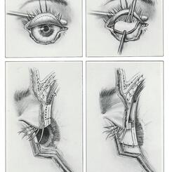
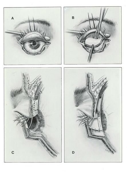 Surgical repair of dog bite to eyelidThis carbon dust on ledger paper illustration depicts the surgical repair of the lower eyelid.
 -
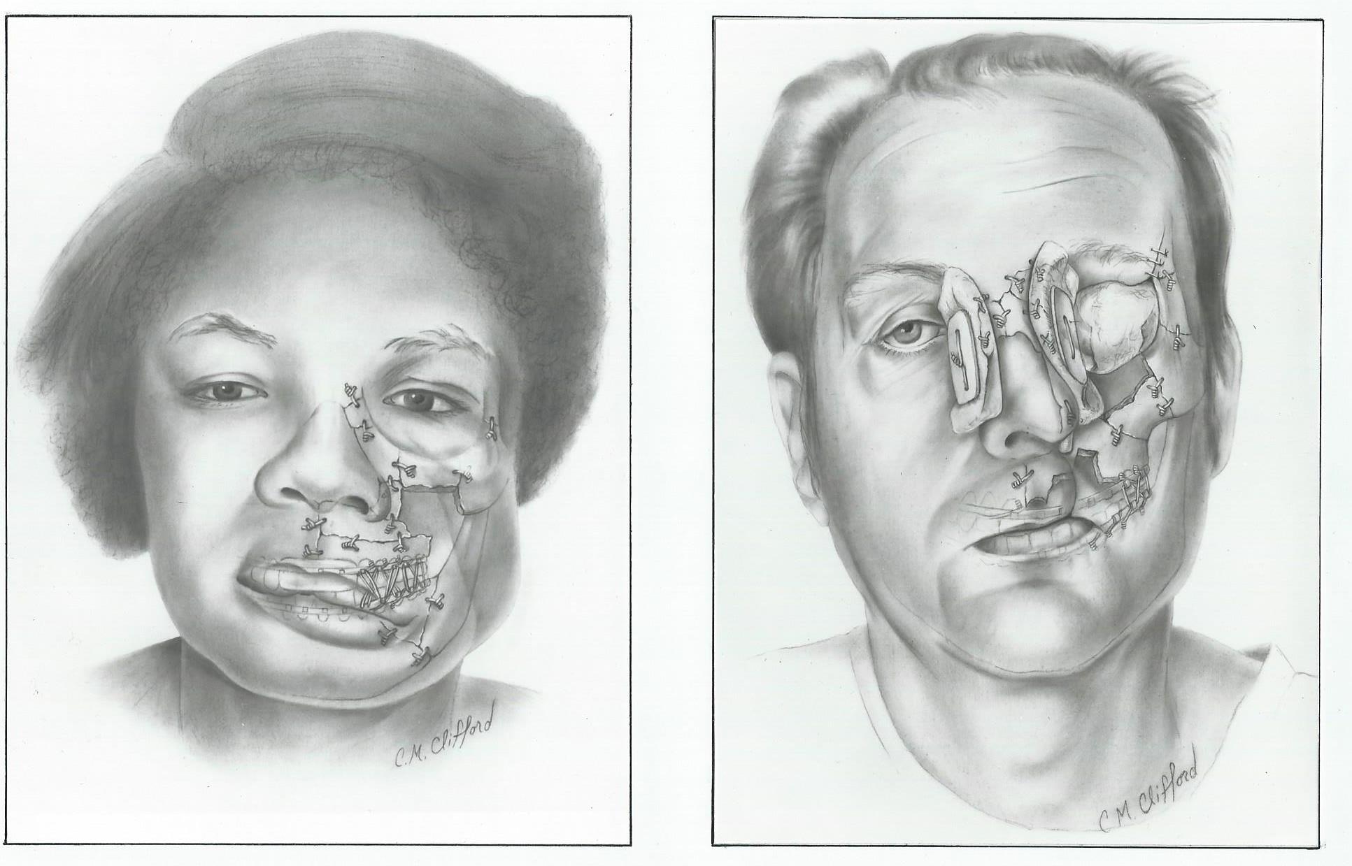
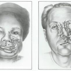
 Gun shot wounds to the faceThese carbon dust illustrations on calcium board depict internal fixation of fractured bones, external fixation with splints, and how the soft tissue on the skull conforms to the underlying structure.
 -
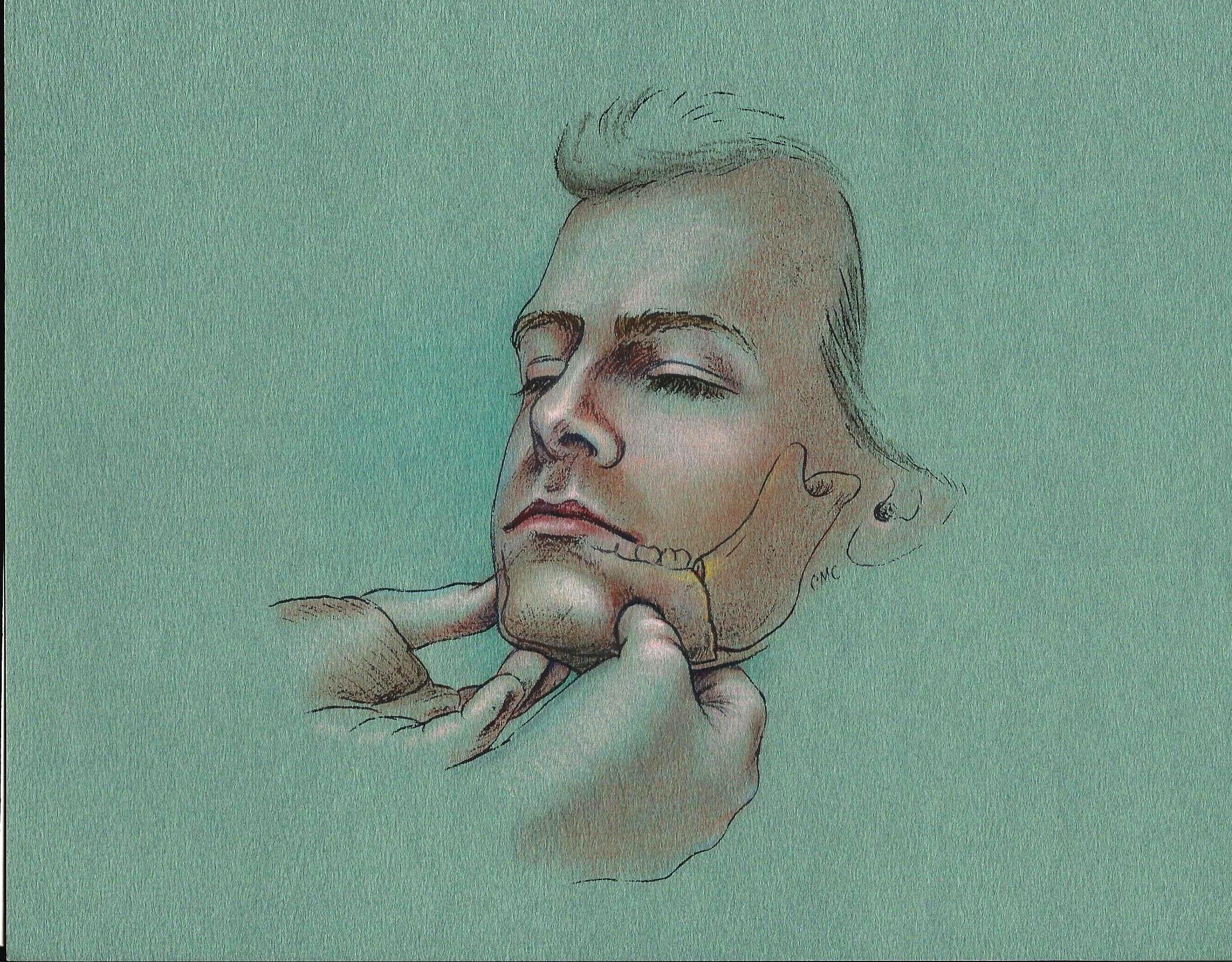
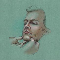
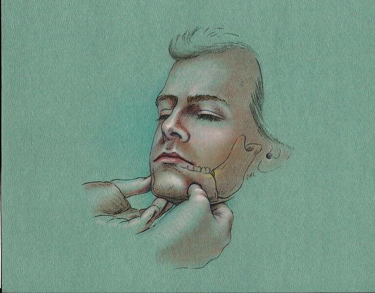 Palpating for fractured jawMixed media on mi tientes paper. The surgeon used this slide in a presentation to teach patient examination techniques.
 -
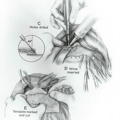
 Surgical repair of fractured frontal bone of foreheadMiddle plate of a series designed to teach surgical techniques. I sketched these in the operating room and rendered them later in my office. Carbon dust on calcium board.
 -
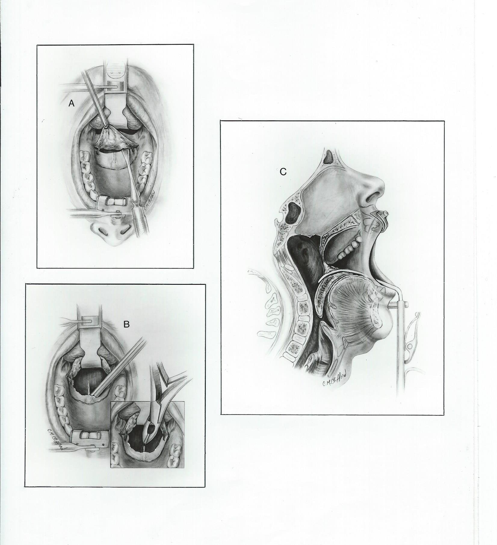
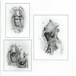
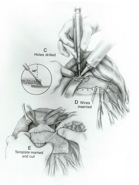
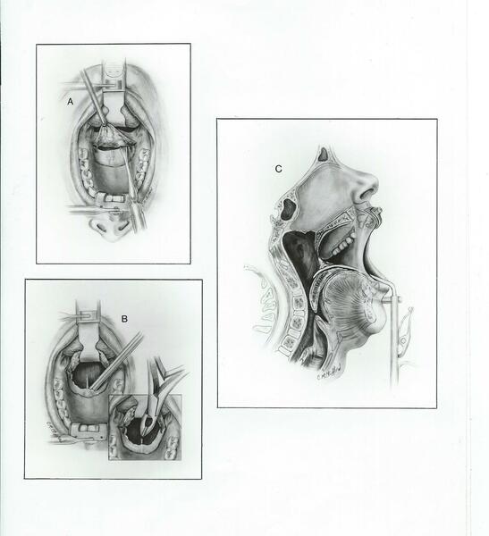 Transpalatal Approach to the NasopharynxCarbon dust on ledger paper. I used a co-worker model for the profile and nose drawings. 9x12 inch sheets.
 -
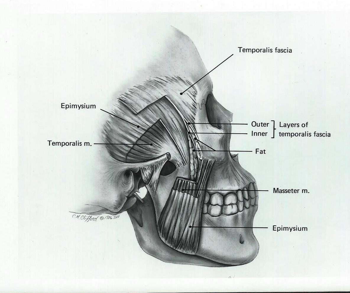
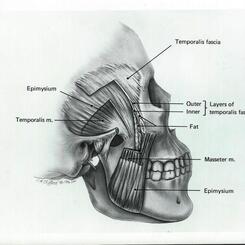
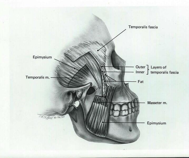 Fascial layersCarbon dust on calcium board. Using the same skull as before and my knowledge of anatomy I created this image to depict these surgically important layers.
 -
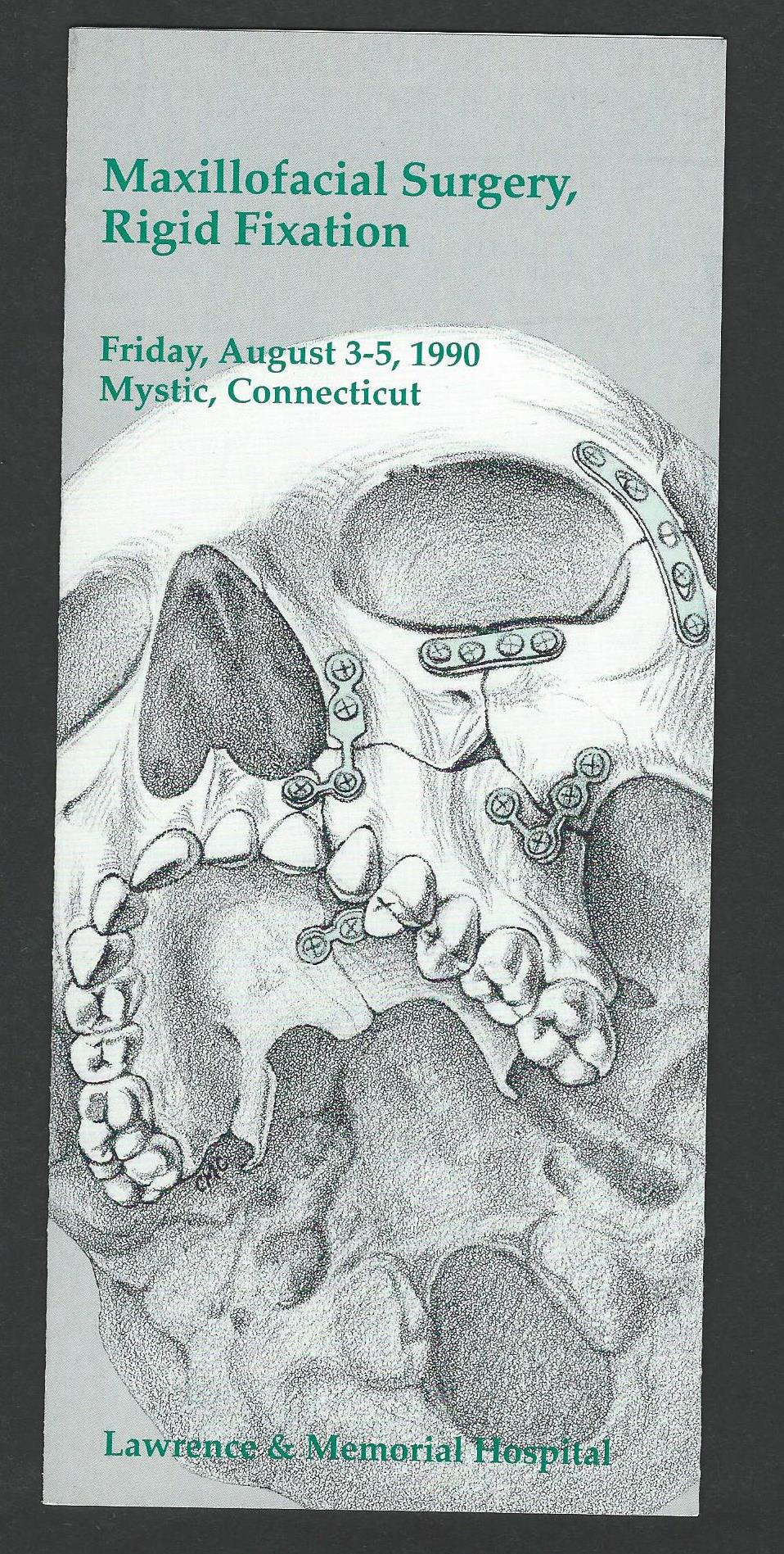
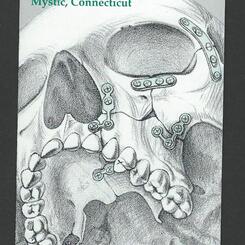
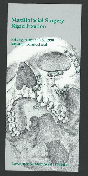 Fracture repair of the maxilla and zygomaBlack pencil on coquille board. This was the last of a series of brochures I illustrated and designed for the Rigid Fixation Conference.